

The short answer
High ALT or ALP is a clue—not a diagnosis.
ALT usually increases when liver cells are irritated or injured. ALP commonly rises when bile flow is affected or the enzyme is induced by hormones or medicines. Either can be raised for several reasons, including conditions that do not begin in the liver.
Your vet will consider the pattern, degree and duration of the changes alongside other blood values and your dog’s health. Some dogs need only a planned recheck; others benefit from urine tests, functional testing, abdominal ultrasound or carefully selected sampling.
What are liver enzymes?
Enzymes are proteins involved in chemical processes within cells. A blood test measures their activity in the circulation. When liver cells are injured, bile flow is disturbed or enzyme production is stimulated, certain values can increase.
“Liver enzymes” and “liver function” are not the same. Enzymes can be high while the liver still performs its essential jobs, and significant liver disease can occasionally exist without dramatic enzyme increases. Measures such as bilirubin, albumin, glucose, urea, cholesterol, bile acids and clotting tests may add information about function, depending on the case.
MSD Veterinary Manual: Overview of hepatic disease
- ALT — alanine aminotransferase
- Primarily a marker of liver-cell injury in dogs. It is sensitive, but it does not reveal the cause, exact severity or remaining liver function.
- ALP — alkaline phosphatase
- May rise with cholestasis, gallbladder or bile-duct disease, steroid exposure, Cushing’s syndrome, some medicines and normal bone growth in young dogs.
- AST — aspartate aminotransferase
- Can increase with liver injury but is also present in muscle and other tissues, so it is interpreted with ALT and sometimes creatine kinase.
- GGT — gamma-glutamyl transferase
- Can support concern about bile-duct or cholestatic disease but, like ALP, is not a stand-alone diagnosis.
- Bilirubin
- A yellow pigment rather than an enzyme. A rise may occur with red-cell breakdown, liver dysfunction or impaired bile flow and can cause jaundice.
Does the size of the increase tell us how serious it is?
Not reliably on its own. A marked result deserves attention, but the highest number is not automatically the most advanced disease. ALT reflects leakage from injured cells rather than the amount of healthy liver remaining. ALP is particularly sensitive in dogs and can rise substantially because of steroid hormones or certain medicines.
Your vet will look at:
- Which values are raised and their relative pattern
- Whether results are improving, stable or rising on repeat tests
- Your dog’s age, breed, symptoms, weight and examination
- All prescribed medicines, supplements and possible toxin exposures
- Bilirubin, proteins, glucose, cholesterol, blood count and urine findings
- Evidence of endocrine, gastrointestinal, pancreatic or other systemic disease
Reference ranges also differ between laboratories. Avoid comparing a current number with an online chart or another dog’s result without the units and laboratory interval.
Why might ALT or ALP be raised in a dog?
Possible explanations range from temporary enzyme induction to primary liver disease. Common diagnostic categories include:
Medicines and hormone effects
Corticosteroids, some anti-seizure medicines and other drugs can alter liver enzymes. Cushing’s syndrome can produce a similar hormonal effect. Never stop essential medication without veterinary guidance; an accurate drug history helps your vet interpret the pattern.
Reactive changes from illness elsewhere
The liver processes blood arriving from the gastrointestinal tract and responds to inflammation throughout the body. Gastrointestinal, pancreatic, endocrine, infectious and other systemic diseases can therefore cause secondary or “reactive” liver changes.
Gallbladder, bile-duct or pancreatic disease
Inflammation, obstruction, gallbladder mucoceles and disease near the bile duct can interfere with bile flow. Bilirubin, ALP and GGT may be particularly relevant, although the precise pattern varies.
Primary liver inflammation or injury
Chronic hepatitis, infection, toxins and copper-associated disease are among the possibilities. Determining the precise type may require histopathology and other laboratory analysis of properly obtained liver tissue rather than blood tests alone.
Nodules, cysts or tumours
Benign age-related nodules and malignant disease can both alter liver appearance or enzymes. Ultrasound helps assess structure, but imaging cannot always distinguish benign from malignant tissue without sampling.
Congenital vascular abnormalities
Portosystemic shunts and some other vascular disorders can affect liver development and function, particularly in younger dogs. The investigation may involve bile acids, ammonia, urinalysis and specialist imaging depending on the presentation.
What tests might my vet recommend next?
The correct next step depends on why the blood test was performed and whether your dog is well. Investigation is usually staged so each result answers a useful question rather than automatically ordering every available test.
- Review the context: symptoms, diet, medication, supplements, toxin risks and previous results
- Repeat or extend blood tests: confirm persistence and review the full biochemical and haematological pattern
- Urinalysis: adds information about hydration, infection, bilirubin, glucose and kidney function
- Functional or targeted tests: bile acids, ammonia, endocrine testing or infectious-disease tests when indicated
- Diagnostic imaging: abdominal ultrasound, and occasionally radiographs or more advanced imaging
- Sampling: selected only when the likely result could change treatment or prognosis
When can an abdominal ultrasound help?
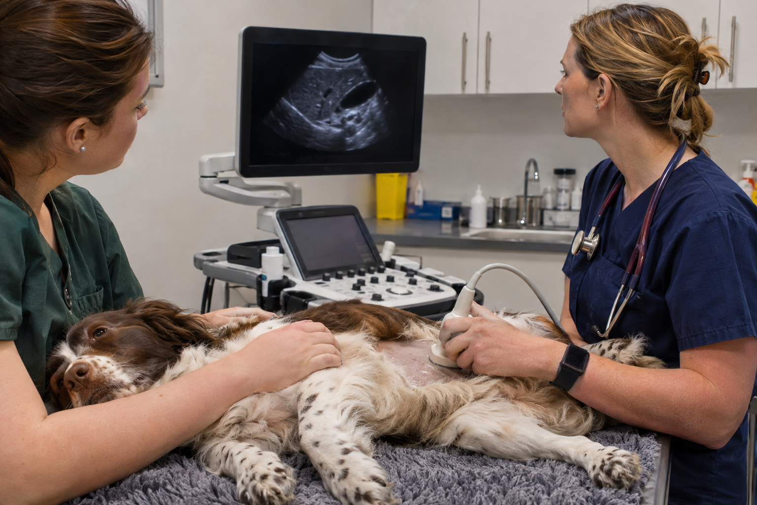
Ultrasound provides a real-time assessment of abdominal structure. It may be recommended when liver abnormalities persist, the dog has relevant symptoms, bilirubin is raised, several tests point towards hepatobiliary disease or your vet needs to investigate another abdominal cause.
An examination can assess:
- Liver size, contour and internal appearance
- Focal nodules, masses, cysts or mineralisation
- The gallbladder, bile ducts and evidence of impaired bile flow
- Blood vessels and selected congenital abnormalities
- The pancreas, gastrointestinal tract, lymph nodes and other abdominal organs
- Free abdominal fluid and safe potential routes for sampling
A normal-looking ultrasound does not exclude every liver disorder. Many inflammatory, metabolic and early microscopic diseases require blood-test interpretation or tissue analysis. Equally, a structural change on ultrasound is not automatically cancer.
What is the difference between a fine-needle aspirate and a core biopsy?
A fine-needle aspirate (FNA) collects cells for cytology. It is less invasive and can help in selected focal or diffuse changes, but it provides limited information about tissue architecture and cannot definitively diagnose disorders such as chronic hepatitis.
A core biopsy collects a larger cylinder of tissue for histopathology. It may provide information about inflammation, fibrosis, copper accumulation and tissue structure. However, it carries greater bleeding and anaesthetic considerations and requires careful planning, clotting assessment, appropriate sample handling and post-procedure monitoring.
Ultrasound guidance can target lesions and help select a safe route, but it does not remove sampling risk. In suspected chronic hepatitis, consensus guidance favours adequate tissue from more than one area and may favour laparoscopic sampling in some patients. The method should be chosen for the individual dog and the diagnostic question.
Available through veterinary referral
A staged liver and internal-medicine workup for suitable dogs.
For dogs referred by their usual veterinary practice, Fabrizio Tucciarone provides postgraduate-qualified internal-medicine and imaging support. Existing results are reviewed first so the next investigation addresses a clear clinical question.
Internal-medicine assessment
History, symptoms, medication, examination and previous laboratory trends are brought together to prioritise hepatic and non-hepatic explanations.
Abdominal ultrasonography
A structured examination assesses the liver, biliary system, pancreas, gastrointestinal tract and other abdominal organs that may explain the pattern.
Image-guided sampling
Fine-needle aspiration or core biopsy may be considered when appropriate, after reviewing bleeding risk, sample requirements and whether the result will change care.
Referral investigations support—not replace—the relationship with your usual veterinary practice. Findings and recommendations are returned to your vet for coordinated ongoing care.
Common questions
High liver enzymes in dogs: FAQs
Do high liver enzymes mean my dog has liver failure?
No. Liver enzymes are mainly markers of cell injury, bile-flow disturbance or enzyme induction; they do not directly measure how well the liver is functioning. Your vet interprets them with symptoms, other blood values, medication, examination findings and changes over time.
What is the difference between ALT and ALP in dogs?
ALT commonly rises when liver cells are irritated or injured. ALP can rise with reduced bile flow, gallbladder or biliary disease, steroid exposure, Cushing’s syndrome and several other situations. Neither result identifies one specific disease by itself.
How high is too high for a dog’s liver enzymes?
There is no single number that determines urgency for every dog. The size and pattern of the increase, whether it persists, the dog’s age and medication, other blood results and the presence of symptoms all matter. A mildly abnormal result in an unwell dog can be more important than a larger isolated result in a dog that appears well.
Can steroids increase ALP in dogs?
Yes. Corticosteroid medicines and increased natural steroid production, as can occur with Cushing’s syndrome, may substantially increase ALP in dogs. Never stop prescribed steroids suddenly; tell your vet exactly what your dog takes and follow their advice.
Can a dog have high liver enzymes without symptoms?
Yes. Abnormal values are sometimes found on pre-anaesthetic, screening or monitoring blood tests before the dog shows obvious signs. Persistent or unexplained changes still deserve interpretation and, where appropriate, a staged investigation.
When is an ultrasound useful for high liver enzymes?
Ultrasound may be useful when abnormalities persist, symptoms are present, several blood markers are affected or your vet suspects liver, gallbladder, bile-duct, pancreatic or other abdominal disease. It can identify structural changes but cannot diagnose every microscopic liver disorder.
Will my dog need a liver biopsy?
Not every dog does. Sampling is considered only when the likely benefit justifies the risks and the result would change treatment. Fine-needle aspiration and core biopsy answer different questions; suspected chronic hepatitis generally requires histopathology from appropriately collected biopsy samples.
Can I book a liver ultrasound directly with Mobile Vet Referral?
Please speak to your usual veterinary practice first. Your vet can interpret the blood results in context, assess urgency and arrange referral for internal-medicine review, abdominal ultrasound or sampling when appropriate.